Figure 5.
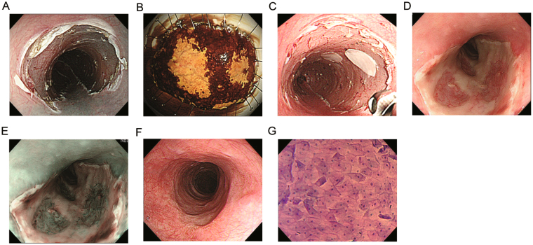
In case #1, semicircular endoscopic submucosal dissection (ESD) spanning seven-eighths of the luminal circumference was successfully performed for middle thoracic oesophageal squamous cell carcinoma (ESCC) (Fig. 5A), and the two ESCC lesions within invasion depth of the lamina propria were both detected by iodine staining (Fig. 5B) in the removed specimen. Six cell sheets were endoscopically transplanted (Fig. 5C). On the 4th day after transplantation, the engrafted cell sheets were present on the post-ESD site (Fig. 5D). NBI-equipped endoscopy enabled distinct identification of the transplants accompanied by plenty of vascularity at the transplantation site with no white exudate (Fig. 5E). Twenty-eight days later, complete ulcer healing with epithelialization was achieved without luminal stricture (Fig. 5F). The transplanted sites were densely covered with regular-shaped squamous cells when observed using endocytoscopy (Fig. 5G). The nuclei of the cells showed nominal abnormality in size and configuration.

