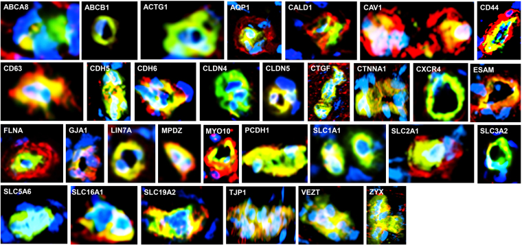
Figure 3.
Validation of the human BNB transcriptome. Merged digital photomicrographs of sural nerve endoneurial microvessels (UEA-1 FITC-positive, green) in situ show BNB endothelial cell expression (yellow-green, yellow or orange co-localization dependent of the relative fluorescent intensity of protein marker in red) of transporters (ABCA8, ABCB1, AQP1, SLC1A1, SLC2A1, SLC3A2, SLC5A6, SLC16A1 and SLC19A2), cytoskeletal proteins (ACTG1, CALD1, FLNA, MYO10), cell membrane proteins (CAV1, CD44, CD63, ESAM), junctional complex proteins (CDH5, CDH6, CLDN4, CLDN5, CTNNA1, GJA1, LIN7A, MPDZ, PCDH1, TJP1, VEZT, ZYX), vascular endothelial cell secreted protein CTGF, and chemokine receptor CXCR4. Positive expression of AQP1, CALD1, CAV1, CD44, CD63, FLNA, MYO10 and SLC2A1 (red) by cells that surround and are in direct contact with endothelial cells (most likely pericytes), and ABCA8 by cells close to but not in direct contact with endothelial microvessels (most likely Schwann cells) are also observed. Nuclei are identified in blue. Original magnification 400X.

