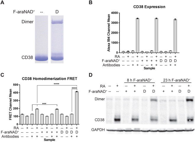
Figure 2.
dF-araNAD+ induces CD38 homodimerization. (A) In vitro labelling of purified CD38. 5 μM purified CD38 was treated with dF-araNAD+ for 30 min as indicated. Reaction mixtures were resolved by SDS-PAGE and stained with Coomassie blue. (B) Mean fluorescence of HL-60 cells stained with Alexa Fluor 594-conjugated CD38 antibody (n = 3). HL-60 cells were cultured for 24 h with 1 μM RA as indicated. At 23 h, cells were treated with 1 μM F-araNAD+ (+) or dF-araNAD+ (D) as indicated. 1 × 10⁶ cells were put in PBS with Alexa Fluor 594-conjugated CD38 antibody as indicated and analysed by flow cytometry. Error bars indicate standard error of the mean (SEM). (C) FRET means (n = 3). HL-60 cells were cultured for 24 h with 1 μM RA as indicated. At 23 h, cells were treated with 1 μM F-araNAD+ (+) or dF-araNAD+ (D) as indicated. 1 × 10⁶ cells were put in PBS with Alexa Fluor 488- and 594-conjugated CD38 antibodies as indicated and analysed by flow cytometry. Error bars indicate SEM. ***p < 0.001; ****p < 0.0001. Two-way analysis of variance (ANOVA) using Tukey’s multiple comparisons test was used to determine significance. (D) Western blot showing covalent dimerization of CD38 when treated with dF-araNAD+. HL-60 cells were cultured with RA and treated with F-araNAD+ or dF-araNAD+ at 8 or 23 h as indicated and lysate was collected at 24 h. 25 μg of lysate per lane was run. Membrane images for each protein are cropped to show only the bands of interest.

