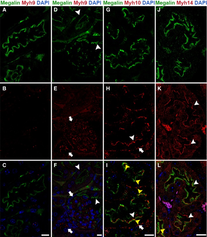
Figure 1.

Myh10 and Myh14 are the NM2 isoforms expressed in the proximal tubular segment. Confocal fluorescence microscopy of the adult mouse kidney sections stained for megalin (proximal tubule marker) and one of the NM2 isoforms revealed that Myh10 and Myh14 are the prominent isoforms of NM2 in the proximal tubules. Myh9 (red) is not expressed in the megalin (green)‐positive proximal tubules (arrowheads, A–F); however, Myh9 expression is present in the adjacent tubules and the glomerulus (arrows, D–F). Myh10 localized to apical (arrowhead) and basolateral (arrow) membranes in the megalin‐positive proximal tubular segments (G–I). Myh14 was expressed in the megalin‐positive tubules on the apical membrane (arrowhead); punctate structures were also observed on the basolateral membrane and within the epithelial cells (J–L). The majority of Myh10 and Myh14 does not colocalize with megalin on the apical membrane, and appear to be above the megalin staining on the membrane. However, there are a few regions (microdomains) on the apical membrane (yellow arrowhead, I&L) where we observe partial colocalization. Sections were mounted with vectashield containing DAPI to stain the nuclei (blue; C, F, I & L). Scale bar = 10 μm.
