Figure 4.

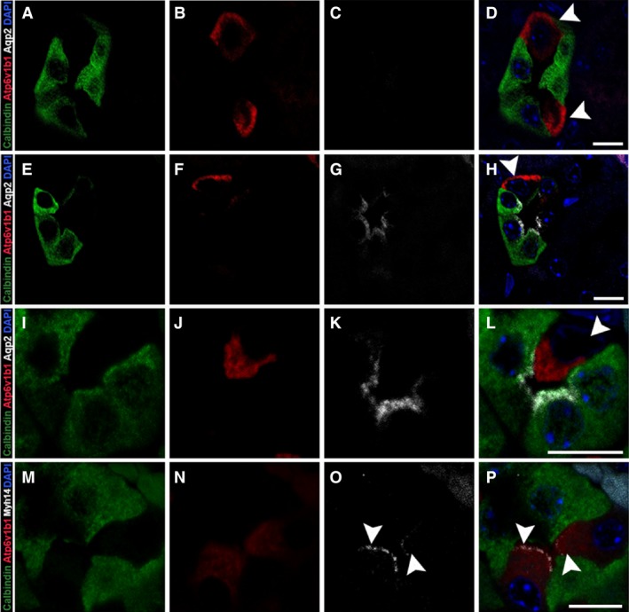
Myh14 localizes to apical membranes of Atp6v1b1‐positive intercalated cells in the distal tubule. Adult kidney sections were stained for calbindin‐D28K, Atp6v1b1, Aqp2, and Myh14 to identify intercalated cells within the distal and connecting tubule segments as well as to confirm Myh14 localization to the intercalated cell type. Cells with positive staining for Atp6v1b1 (red) can be found in tubule segments positive for calbindin‐D28K only (A–D) as well as in tubules positive for both calbindin‐D28K and Aqp2 (E–H); in both cases Atp6v1b1 staining is observed along the basolateral membrane indicating β‐ intercalated cell type (arrowhead, D&H). We also observed Atp6v1b1 staining in majority of the tubules along the apical membrane indicating the α‐ intercalated cell type (arrowhead, I–L). Myh14 localized to the apical membrane of the intercalated cell type in the distal tubule (arrowhead, M–P). Sections were mounted with vectashield containing DAPI to stain the nucleus (blue; D, H, L and P). Scale bar = 10 μm.
