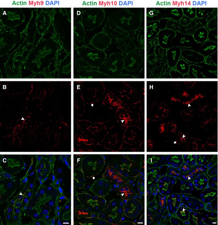
Figure 6.

NM2 isoforms have selective expression and localization pattern with actin filaments in the renal tubules. Confocal analysis of sections stained for phalloidin and NM2 isoforms showed that NM2 isoforms did not colocalize with all the filamentous actin structures in the tubules. Myh9 colocalized with actin filaments on the apical membrane (A–C, arrowhead). Myh10 (D–F) and Myh14 (G–I) colocalized with actin filaments on both the apical (arrowhead) and basolateral membranes (arrow). NM2 isoforms appeared as punctate structures or short filaments on the membrane. Sections were mounted with vectashield containing DAPI to stain the nucleus (blue; C, F & I). Scale bar = 10 μm.
