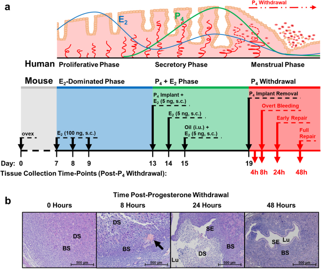
Figure 1.
Schematic comparison of human menstrual cycle (top) and simulated menstruation protocol in an ovariectomised mouse model (bottom). (a) Upper panel depicts a schematic of the endometrium and its changes through the menstrual cycle consequent upon circulating concentrations of oestradiol (E2) and progesterone (P4). Lower panel depicts a schematic of the simulated menses protocol in the ovariectomised mouse model and the time-points at which tissues were collected. ovex = ovariectomy, s.c. = subcutaneous, i.u. = intrauterine. (b) Representative H&E photomicrographs of mouse endometrial tissue sections through decidualisation (0 h; stromal decidualisation, but no tissue breakdown), overt menses (8 h; loss of stromal compartment structural integrity), early repair (24 h; early re-epithelialisation and dissociation of necrotic tissue from basal endometrium) and full repair (48 h; stromal restoration and complete re-epithelialisation) following withdrawal of progesterone. Black arrow indicates active bleeding at 8 h, which can be seen both micro- and macroscopically. BS = basal stroma, DS = decidualised stroma, Lu = lumen, SE = surface epithelium.

