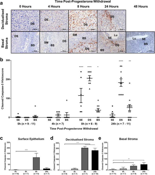
Figure 3.
Apoptosis is significantly increased by 8 hours after progesterone withdrawal in the mouse endometrium. Expression of CC3 was analysed by immunohistochemistry and semi-quantitative histoscoring. (a) Representative photomicrographs of endometrial tissue sections at high power, centred on decidualised stroma and basal stroma. BS = basal stroma, DS = decidualised stroma, Lu = lumen, SE = surface epithelium, SM = smooth muscle. (b) CC3 expression in the surface epithelium (SE; black squares), decidualised stroma (DS; black triangles) and basal stroma (BS; black circles) plotted against time post-progesterone-withdrawal. (c–e) Expression in individual tissues plotted against time post-progesterone withdrawal. Results are presented as mean ± SEM. Significance determined by 1-way ANOVA and Dunnett’s multiple comparisons test (to 0 hours’ progesterone withdrawal). *p < 0.05, **p < 0.01, ****p < 0.0001.

