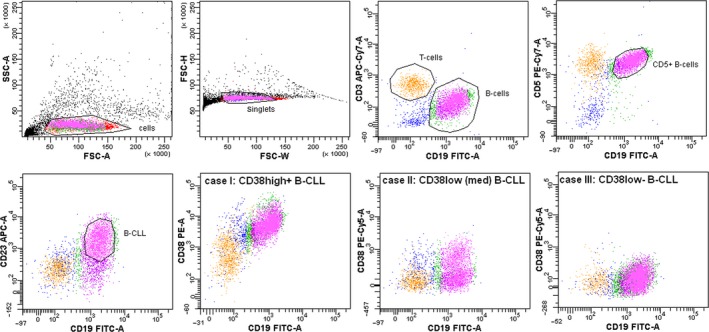
Figure 2.

Flow cytometry and cell sorting strategy for B‐CLL cells. “Cells” are gated from debris according to forward and side light scatter characteristics; “singlets” are gated according to the FSC signal width/height ratio; “B‐cells” are gated based on CD19 fluorescence; B‐CLL cells are further gated based on the CD3‐CD19+CD5+CD23+ cell phenotype; the gating is sequential. The lower panel presents three different cases of B‐CLL with varying densities of surface CD38 (identified as high+, low [med], and low−).
