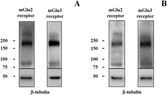
Figure 5.

Western blot analysis reveals the presence of mGlu2 receptor and mGlu3 receptor protein dimers in mouse cortical (A) and spinal cord (B) synaptosomes (10 μg protein for cortical lysates and 20 μg protein for spinal cord lysates). The figure shows a representative blot of five (cortex) to seven (spinal cord) analyses carried out on different days.
