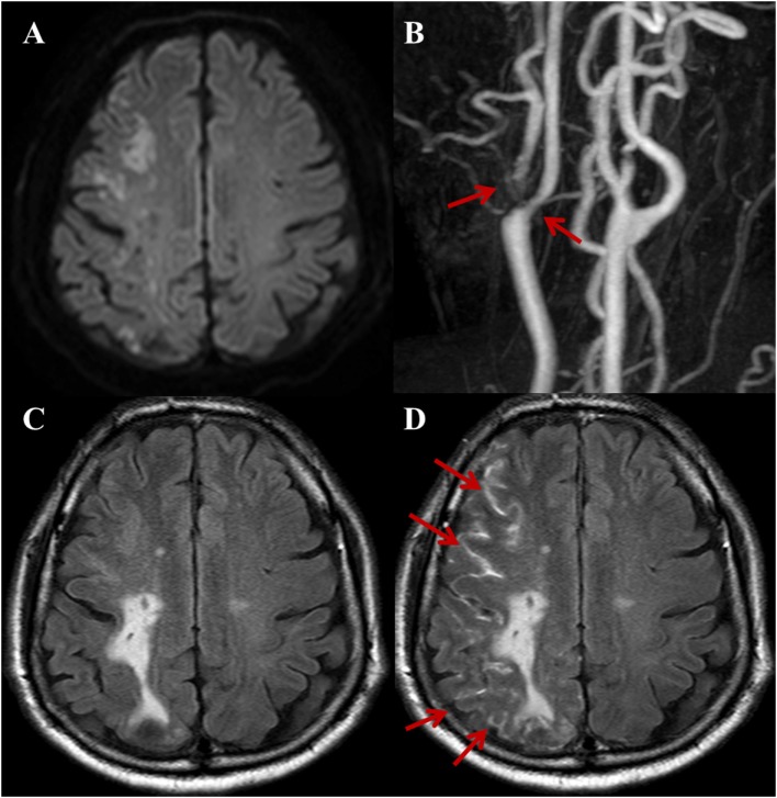
Figure 1.
A 72-year-old male with a complaint of acute onset left arm weakness. Stroke due to large artery to artery atherosclerosis. (A) DWI shows acute border zone infarctions on the right cerebral hemisphere. (B) Three-dimensional time-of-flight MRA of extracranial arteries shows moderate-to-severe stenosis with plaque at right proximal internal carotid artery and ECA (arrows). Compared with non-contrast fluid-attenuated inversion recovery (FLAIR) (C), the post-contrast FLAIR (D) image shows diffuse sulcal enhancement (hyperintense acute reperfusion marker sign) along the right cerebral sulci (arrows).

