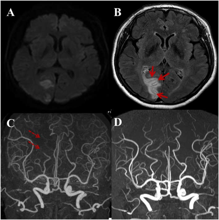
Figure 4.
A 59-year-old male with a complaint of acute onset left side visual field defect in both eyes. Regional pattern of hyperintense acute reperfusion marker. (A) DWI shows acute infarction at the right occipital lobe involving the visual cortex. (B) Post-contrast fluid-attenuated inversion recovery image shows sulcal enhancement along the right occipital lobe (arrows). Compared with outside CTA (C), three-dimensional time-of-flight MRA (D) shows the recanalization state of right distal posterior cerebral artery (dashed arrows).

