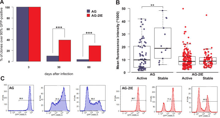
Figure 2.
Stability of expression and silencing of provirus transcription in single-cell clones. (A) The percentages of stable clones with ≥ 90% of GFP-positive cells 3, 30 and 60 dpi are shown for AG and AG-2IE virus vectors. Three dpi all clone-establishing cells were GFP-positive. In total, 2,128 and 558 clones with AG and AG-IE proviruses were established, respectively. Thirty dpi 10% of AG (210) and 39% of AG-2IE (218) clones kept stable expression of GFP. Sixty dpi 3.5% (74) and 29% (174) of AG and AG-2IE clones kept stable expression. (B) Mean fluorescence intensities (MFI) of AG and AG-2IE clones. Each dot represents one clone. Clones were established by sorting GFP-positive cells. MFIs of the clones with at least 20% GFP-positive cells at 21 dpi (active) and clones with at least 90% GFP-positive cells (stable) at 30 dpi were compared. (C) Histograms of GFP expression of representative clones. Silenced clones of AG show gradual decrease of GFP intensity, while AG-2IE clones silence with a bimodal profile.

