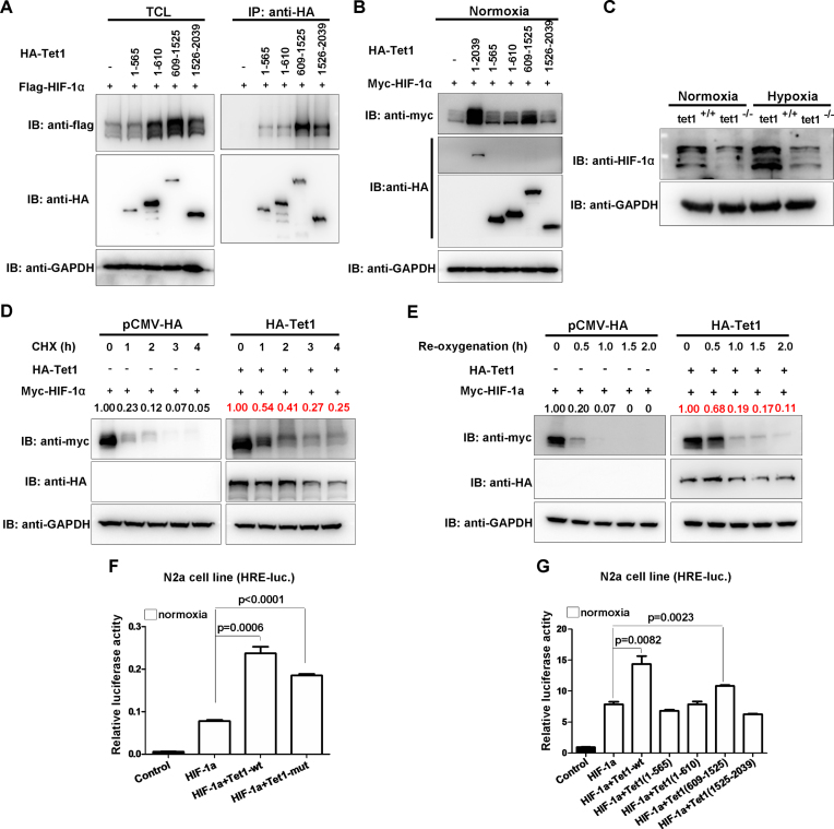
Figure 6.
Tet1 stabilizes HIF-1α protein and enhances HIF-1α transcriptional activity independent of its enzymatic activity. (A) Domain mapping of Tet1 protein binding to HIF-1α. (B) Tet1 and its different domains stabilized HIF-1α protein. (C) The protein level of HIF-1α was reduced in Tet1-null MEF cells compared with that in wild-type MEF cells under normoxia and hypoxia. (D) Overexpression of Tet1 enhanced stabilization of HIF-1α in the presence of cycloheximide (CHX, 50μg/mL) under normoxia. HEK293T cells were transfected with the indicated plasmids for 18-20 h, then, cycloheximide was added to culture medium with the final concentration 50μg/mL. At different time points, the cells were harvested for Western blot analysis. (E) Overexpression Tet1 enhanced stabilization of HIF-1α during re-oxygenation. HEK293T cells were transfected with the indicated plasmids for 16-18 h. Subsequently, the cells were placed in a hypoxia incubator (NBS Galaxy 48R) for 8 h (5% O2). Then, the cells were transferred into a regular incubator (21% O2). At different time points, the cells were harvested for Western blot analysis. (F) Overexpression of mouse Tet1 (Tet1-wt) and its enzymatic inactive form (Tet1-mut) together with HIF-1α could activate the hypoxia response element luciferase (HRE-luc.) reporter induced by HIF-1α in the N2a cell line under normoxia. (G) Overexpression of mouse Tet1 (Tet1-wt) and its domain covering 609-1525aa together with HIF-1α could activate the HRE-luc. reporter induced by HIF-1α in the N2a cell line under normoxia.

