Figure 2.
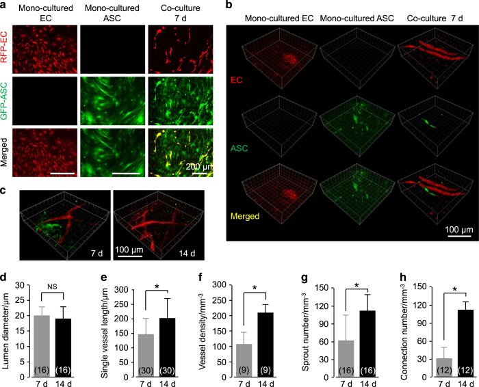
Angiogenesis in a 3D collagen model by co-culturing ASCs and ECs in vitro. (a) Formation of vessel-like structures in ASC-EC co-cultured 3D gels imaged by enhanced inverted microscopy. ASCs are GFP-positive and ECs are RFP-positive. The images shown are representative of four different experiments (n=4). (b and c) Formation of vessel-like structures in ASC-EC co-cultured 3D gels further imaged and reconstructed by modified CLSM. The images shown are representative of four different experiments (n=4). (d–h) Analyses of angiogenesis in ASC-EC co-cultured 3D gels. The numbers shown in parentheses indicate images (vascular-like structure diameter (d) and single vessel length (e)) examined in each case, and similar results were observed in four independent experiments (n=4). Analyses of vessel density (f), sprout number (g), and connection number (h) were performed for at least three independent experiments (n=3). *P<0.05; NS, no significant difference.

