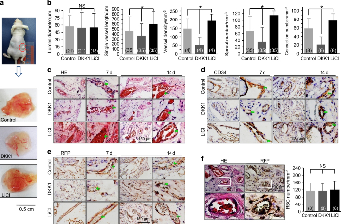
Figure 5.
The in vivo capillary development of the implants is modulated by the Wnt pathway. (a) Representative morphologies of capillaries in the implanted gels (14 days) collected from the subcutaneous pockets at both dorsal sides of nude mice are regulated by DKK1 and LiCl. (b) Analyses of angiogenesis in the implants. The numbers shown in parentheses indicate images (vascular-like structure diameter and single vessel length) examined in each case and similar results were observed for four independent experiments (n=4). The analyses of vessel density, sprout number, and connection number were performed for at least three independent experiments (n=3). *P<0.05; NS, no significant difference. (c) H&E stain showing newly formed capillaries with red blood cells inside. The images shown are representative of four experiments (n=4). (d) Positive immunohistochemical staining of RFP implies that these newly formed functional capillaries in the sections originated from the implanted endothelial cells. The images shown are representative of four experiments (n=4). (e) Positive immunohistochemical stain of CD34 indicates that these newly formed functional capillaries in the sections originated from the implanted cells. The images shown are representative of four experiments (n=4). (f) H&E stain and RFP stain from one implanted gel section indicates that these newly formed functional capillaries originated from the implanted endothelial cells. The images shown are representative of four experiments (n=4).

