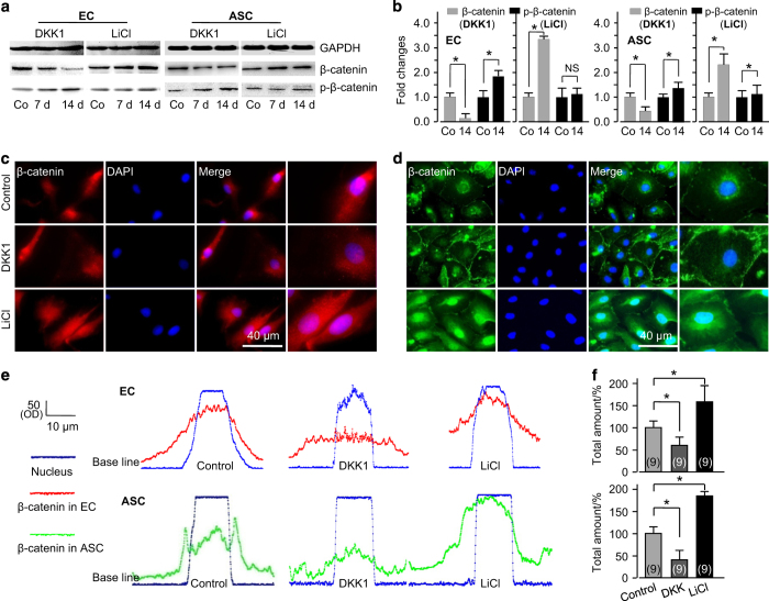
Figure 6.
Wnt-regulated ASC-paracrine angiogenesis is based on nuclear translocation of β-catenin. (a) Western blot showing that β-catenin/phosphorylated β-catenin was regulated by DKK1 and LiCl in both ECs and ASCs. The gels shown are representative of three different experiments (n=3). (b) Quantified analyses of β-catenin/phosphorylated β-catenin regulated by DKK1 and LiCl in ECs and ASCs at 14 days using Quantity One 4.6.3 software. *P<0.05; NS, no significant difference. (c) Immunofluorescent stain showing the nuclear accumulations of β-catenin in ECs and ASCs at 14 days after treatment with LiCl or DKK1. The images shown are representative of three different experiments (n=3). (d) Representative linear fluorescence quantification showing the accumulation of nuclear translocated β-catenin in ECs and ASCs regulated by DKK1 and LiCl. (e and f) Total amounts of β-catenin translocated into the nucleus in both ECs and ASCs. The data shown are representative of nine different experiments (n=9). *Significant difference with respect to the control group (P<0.05).

