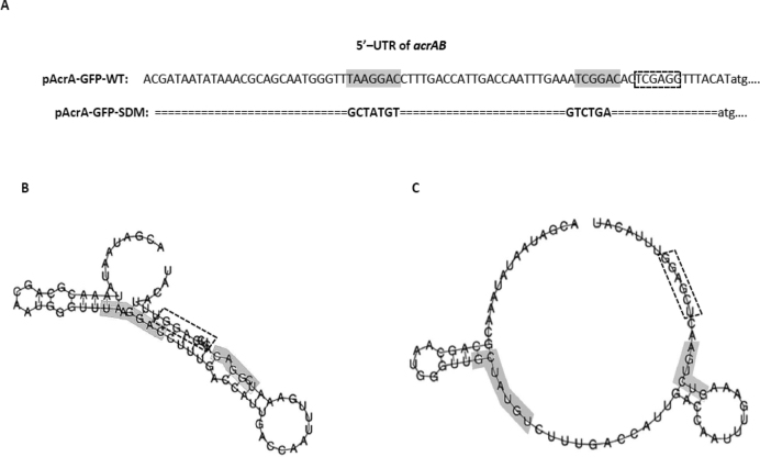
Figure 8.
Primary sequence and secondary RNA structures of WT and mutant acrAB leader regions. Primary DNA sequence of wildtype 5′ UTR of acrAB and mutated 5′ UTR of acrAB in which the nucleotides making up the CsrA binding sites have been substituted (gray shaded box). Ribosome binding site (dashed rectangle box). (A) The RNA structures of the acrAB upstream region in the two plasmids of pAcrA-GFP-WT (B) and pAcrA-GFP-SDM (C) were predicted by RNAfold and their folding free energies were −12.30 kcal/mol and −6.0 kcal/mol, respectively. (Substituted nucleotides in gray shaded box)

