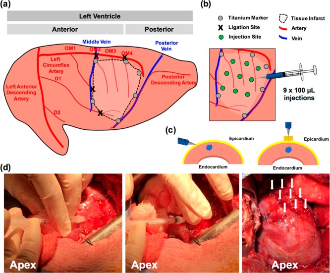
Figure 7.
Intramyocardial hydrogel injection in a pig model of myocardial infarction. (a) Schematic of ligation sites in obtuse marginal branches of the left circumflex artery and diagonal branches of the left anterior descending artery. Titanium markers are placed circumferentially around the infarct for visualization after injection. (b) Traditional injection model for porcine infarct models, consisting of 9 × 100 μL injections. Titanium markers are placed over injection sites prior to hydrogel injection to ensure consistent spacing and to avoid vasculature during injection. (c) Techniques for injection into porcine tissue. Angling the needle parallel to myocardium will allow for penetration directly into myocardium without injection into the ventricle. A foam spacer can also be placed on the needle to permit injection directly into the epicardium without angling the needle. (d) Photos of injection, demonstrating needle position and hydrogel injection into sites marked by titanium markers, finger placement to prevent leak of material after injection as the needle is withdrawn, and 9 injection sites marked by titanium markers following injection into the infarct.

