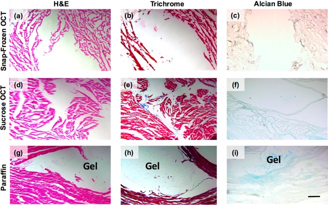
Figure 8.
Hydrogel and tissue preservation after embedding, sectioning, and staining. Ex vivo tissue after being (a–c) snap-frozen in OCT, (d–f) frozen in OCT following sucrose infiltration, or (g–i) embedded in paraffin. (a, d, g) H&E, (b, e, h) Masson’s Trichrome, and (c, f, i) Alcian Blue staining was performed to visualize the hydrogel within the tissue. Scale bar: 200 μm.

