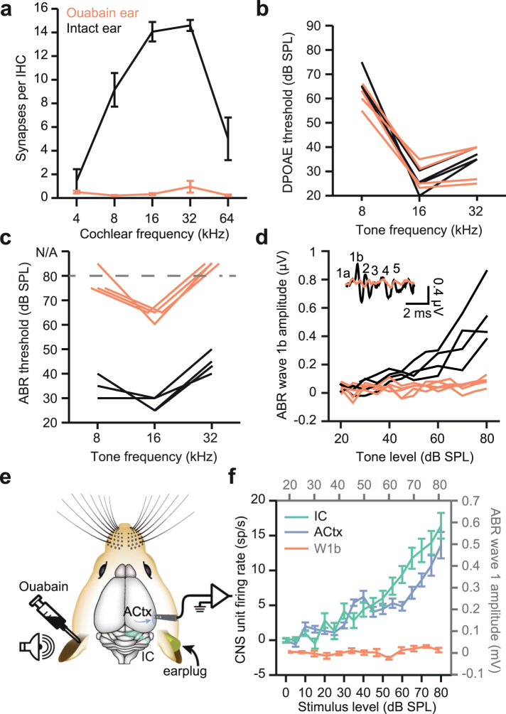
Figure 1.
Ouabain treatment selectively degenerates spiral ganglion neurons, eliminating the ABR without affecting cochlear amplification. (a) Cochlear afferent synapses (operationally defined from juxtapositions of inner hair cell synaptic ribbons and auditory nerve glutamate receptors) were made from immunohistochemically stained sections corresponding to cochlear frequency locations. In ouabain treated ears (n = 4 mice, red line) compared to control (black line), more than 95% of synapses were eliminated by ouabain treatment at least 30 days prior. (b) Hair cell function was preserved 30 days after ouabain treatment, as shown via DPOAE threshold measurements taken at various F2 frequencies. Thresholds from the ouabain-treated left ear (red) and untreated right ear (black) are shown from individual mice after ouabain treatment (n = 4 ears). (c) Auditory brainstem response (ABR) wave 1b threshold measurements were significantly higher from the ouabain-treated ear, indicating a near-complete loss of auditory nerve fibers. (d) ABR wave 1b (inset) amplitudes show a barely detectable or absent signal from the ouabain-treated ear. (e) Schematic of midbrain and cortex recording experiments. Adult mice treated with ouabain 30 days prior were implanted with chronic recording electrodes (see Methods) in the IC and ACtx contralateral to the ouabain-treated (left) ear. (f) Although ABR measurements indicated a near-complete loss of activity in the auditory nerve, central stations of the auditory pathway showed robust responsiveness to sound and monotonically increasing rate-level functions (green line = IC; blue line = ACtx). All data in Fig. 1 have been published in an earlier report12.

