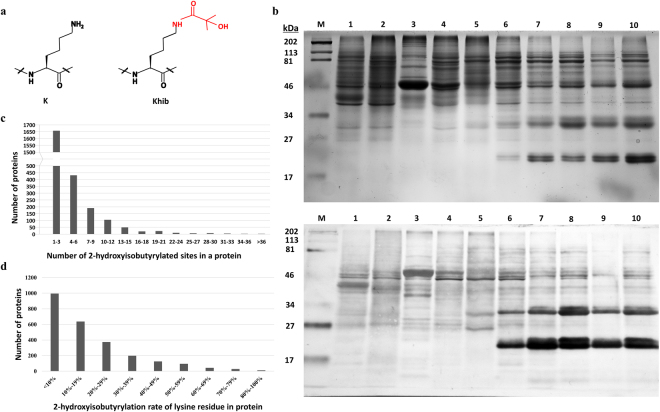
Figure 1.
Lysine 2-hydroxyisobutyrylation in different rice tissues and proteins. (a) Structure of lysine 2-hydroxyisobutyrylation. (b) Lysine 2-hydroxyisobutyrylation profile in different rice organs/tissue revealed by western blotting. Molecular weight is labelled on the left. The samples are labelled on the top. M: size marker; 1. suspension cell protein; 2: roots protein; 3: leaves protein; 4: flowers protein; 5: pollens protein; 6: protein from 7 dpa seeds; 7: protein from 15 dpa seeds; 8: protein from 21 dpa seeds; 9: protein from mature dry seeds; 10: protein from mature dry seeds. Upper: Image of SDS-PAGE stained with coomassie blue. Lower: western blotting image. Same amount of proteins (25 μg per lane) were loaded for sample 1–8 and 10; 20 μg protein was loaded for sample 9. The original images of SDS-PAGE and western blotting are shown in Supplementary Fig. S1. (c) Distribution of 2-hydroxyisobutyrylated proteins based on number of modification sites. (d) Distribution of 2-hydroxyisobutyrylation rate on lysine residues of 2-hydroxyisobutyl-proteins.

