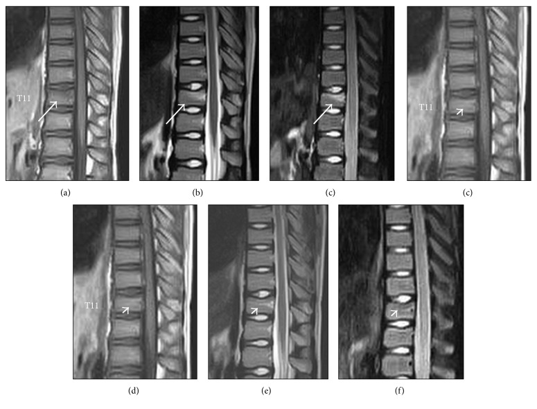
Figure 1.
Magnetic resonance images at the initial visit showing low signal on T1WI (a), relatively high signal on T2WI (b), and high signal on short tau inversion recovery (STIR) image (c) in the upper part of the vertebral body at T11 without deformation of the cortical endplate (arrow). At 1-month follow-up later, signal changes had diminished on T1, T2, and STIR images, respectively (d, e, and f, arrowhead).

