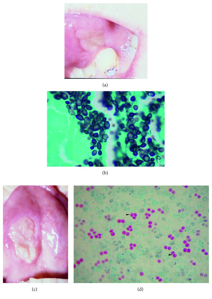
Figure 1.
(a, b) Lesions on the tongue (4 × 2 cm and 3 × 2 cm) with slightly rolled and raised edges. (c) High-power view of a Gomori methenamine silver stain (GMS). Biopsy from ulcerated vocal cord lesion demonstrates spherical to oval yeast cells, some of which are budding. (d) High-power view of a Kinyoun stain. Round ascospores (arrowhead) inside the asci that are characteristic of S. cerevisiae.

