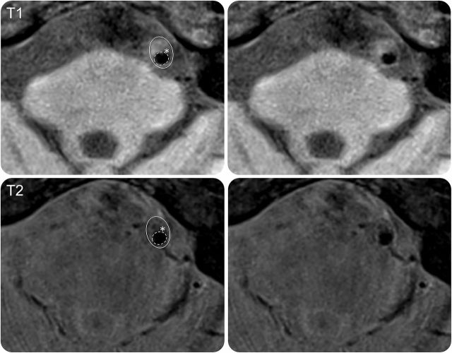
Figure. Proximal basilar artery symptomatic stenosis.
Image shows a cross-section of a symptomatic basilar artery plaque. Top row T1 and bottom row T2 images show heterogeneous plaque with lipid (isointense on T1, hypointense on T2) and irregularity of the lumen surface (dashed line) over the plaque consistent with ruptured fibrous cap (white asterisk). Figure courtesy of Dr. Tanya Turan.

