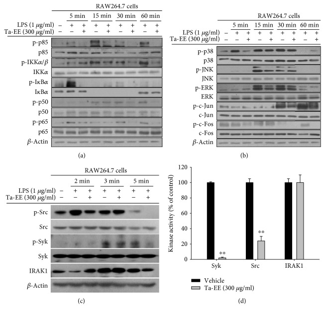
Figure 4.
Ta-EE suppressed the activation of the NF-κB and AP-1 signaling pathways in LPS-stimulated RAW264.7 cells. RAW264.7 cells pretreated with Ta-EE (300 μg/ml) for 30 min were treated with LPS (1 μg/ml) for the indicated time, and the total and phosphorylated protein levels of (a) p85, IKKα/β, IκBα, p50, and p65; (b) p38, JNK, ERK, c-Jun, and c-Fos; and (c) Src, Syk, and IRAK1 in the total cell lysates were determined by Western blot analysis. β-Actin was used as an internal control. (d) Effects of Ta-EE on the kinase activities of Src, Syk, and IRAK1 were determined by in vitro kinase assay using purified Src, Syk, and IRAK1 as described in Materials and Methods. ∗P < 0.05 and ∗∗P < 0.005 versus a control group.

