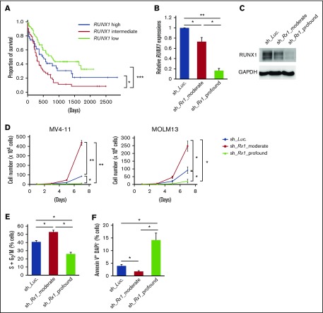
Figure 1.
Moderate inhibition of RUNX1 confers proliferative advantage to AML cells. (A) Overall survival of AML patients from TCGA clinical datasets (n = 187). Patients were divided into 3 groups according to their RUNX1 expressions (RUNX1 high: n = 62; RUNX1 intermediate: n = 63; RUNX1 low: n = 62). (B) Efficacy of shRNAs targeting RUNX1. MV4-11 cells were transduced with lentivirus encoding shRNA targeting Luciferase (sh_Luc.) or shRNAs against RUNX1 (sh_Rx1_moderate 1 or sh_Rx1_profound 1) and incubated with 3 μM of doxycycline for 48 h, then total RNA was prepared and analyzed by RT-PCR. Values are normalized to that of control vector-transduced cells (n = 3). (C) Immunoblot showing the RUNX1 expressions in panel B. (D) Growth curves of MV4-11 cells transduced with control (sh_Luc.) or with RUNX1 shRNAs (sh_Rx1_moderate 1 or sh_Rx1_profound 1). Cells were cultured in the presence of 3 μM of doxycycline (n = 3). (E) RUNX1 depletion-mediated change in the number of cells with S + G2/M phase DNA content. MV4-11 cells transduced with control (sh_Luc.) or with RUNX1 shRNAs (sh_Rx1_moderate 1 or sh_Rx1_profound 1) were cultured in the presence of 3 μM of doxycycline. Forty-eight hours after treatment, cells were harvested and subjected to flow cytometric analysis (n = 3). (F) Frequency of early apoptotic cell death induced by RUNX1 silencing. MV4-11 cells transduced with control (sh_Luc.) or with RUNX1 shRNAs (sh_Rx1_moderate 1 or sh_Rx1_profound 1) were treated as in panel E, and the early apoptotic cells (annexin V+ DAPI−) were scored by flow cytometric analysis (n = 3). Data are mean ± SEM values. *P < .05; **P < .01, by 2-tailed Student t test (except for panel A); ***P < .001, by log-rank (Mantel-Cox) test.

