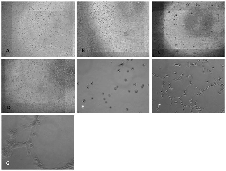
Figure 1.
Representative live cell images taken at two time points. Human chondrocytes (A) and Hela cells (B) are randomly distributed after 1 h of cell culture. After 24 h human chondrocytes have clustered (C), whereas Hela cells (D) do not cluster. Magnification 20× Human chondrocytes in culture after 1 h (E) and after six hours they start forming clusters and develop filopodia/cell surface protrusions to get in contact to each other. These filopodia pull the cells to each other’s. Interestingly there are several cells not being involved in this process not migrating towards each other’s. (F). After 24 h human chondrocytes have formed clusters of several cells. (G) Magnification 40×.

