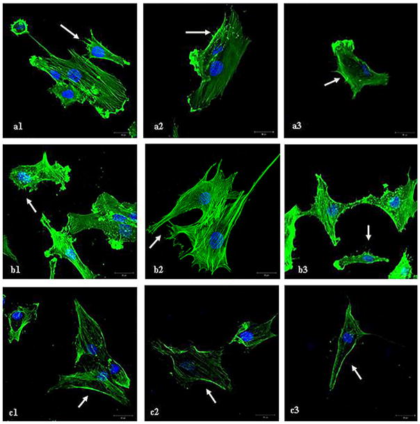
Figure 4.
F-actin staining on human chondrocytes samples from three patients. After treatment with the 2A3 antibody human chondrocytes show a decrease in the appearance of small cellular protrusions on the cell surface (c) compared to untreated human chondrocytes (a) and human chondrocytes treated with a human immunoglobuline a control (b). Microspikes and the changed cell surface are marked with an arrow (→). The reduction was described semiquantitativly (+, ++, +++) from two blinded investigators.

