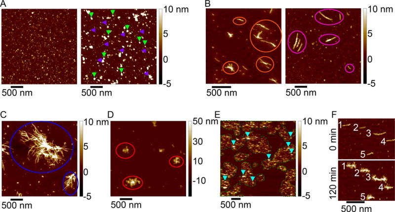
Figure 3. Atomic force microscopy images of a variety of aggregates formed by htt-exon1 proteins.

(A) Htt exon1 can form a variety of globular, oligomeric species (purple arrows indicate oligomers ~5 nm in height; green arrows indicate oligomers ~10 nm in height). (B) Two morphologically distinct fibrils structures formed by exon1, (orange circles indicates thinner, smooth fibril structures; pink circles indicate thicker fibrils with a beaded morphology). (C) Blue circles indicate large bundles of htt exon1 fibrils. (D) Large, amorphous aggregates of htt exon1 are indicated by red arrows (note the color scale goes up to 50 nm). (E) When htt exon1 aggregates on a lipid bilayer, a variety of oligomeric aggregates (blue arrows) associated with regions of increased membrane roughness (outlined with the green dashes lines) are observed. (F) When adding monomeric htt exon1 to pre-formed fibrils, the monomer can accumulate around the fibrils and form a variety of branching points (numbers indicated the same fibril at 0 minutes and 120 minutes after exposure to monomeric htt exon1).
