Fig. 1.

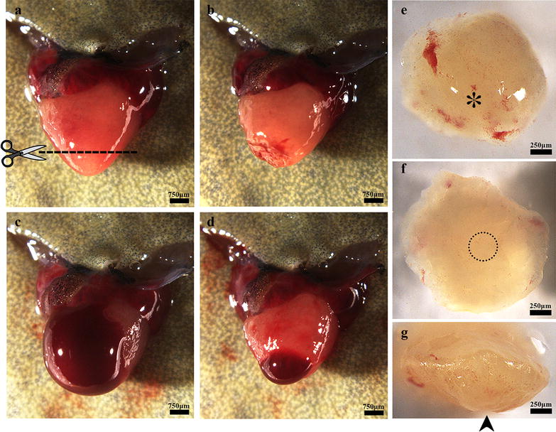
Apical resection of the X. tropicalis heart and rapid blood coagulation after resection. a The heart was exposed for apical resection. The dash line shows the resection plane, corresponding to approximately 10% of the ventricle. b The heart immediate after surgical amputation. c Bleeding after ventricle amputation. d Rapid blood coagulation in the amputated heart after approximately 5 s of pressure with sterile cotton. e Outer surface of the amputated apex. The asterisk indicates the end of the apex on the outer surface. f Inner surface of the amputated apex. The circle with the dotted line indicates the bottom of the ventricle. g Lateral view of the amputated apex. The arrow indicates the bottom of the apex on the outer surface
