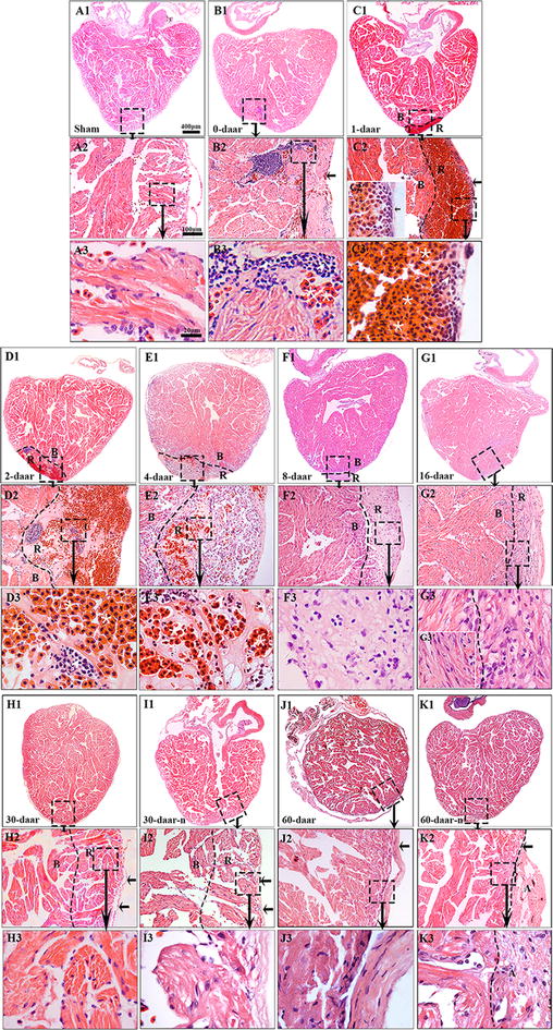
Fig. 3.

Adult X. tropicalis heart has capacity for regeneration after resection. a1 Longitudinal section of an adult heart from the non-amputated sham control. Bar = 400 μm. a2 High magnification of the region outlined by rectangle in a1. Bar = 100 μm. a3 High magnification of the rectangle region in a2. Bar = 20 μm. b1–b3 An amputated heart at 0 daar (approximately 30 min after amputation), showing activated inflammation and hyperemia at the border of the injury. c1–c3 An amputated heart at 1 daar, showing a membrane-like structure (arrow) close to the outer surface of the scar tissue which exhibited with inflammatory cell infiltration, and red blood cells accumulation below the membrane-like structure. c2′ High power of the membrane-like structure which is consisted of cellular or some extracellular matrix. d1–d3 An amputated heart at 2 daar, showing more inflammatory cells in the regenerated area compared with 1 daar. e1–e3 An amputated heart at 4 daar, showing an increase of inflammatory cells and a decrease of red blood cells in the regenerated area compared with 2 daar. f1–f3 An amputated heart at 8 daar, showing the disappearance of most infiltrated red blood cells and a significant decrease of the inflammatory cell intensity in the regenerated area. g1–g3 Most of the fibrous tissue production was replaced with newly regenerated cardiomyocytes and some of the newly regenerated cardiac myocytes are matured, as indicated by a more regular cardiac-specific cross-striated morphology at 16 daar (g3′). h1–h3, i1–i3 An amputated heart regenerated with a perfect morphology and nearly perfect morphology at 30 daar. The amputated area was regenerated with mature cardiomyocytes. In addition, the regenerated myocardium has an intact epicardium (arrow). j1–j3, k1–k3 An amputated heart regenerated with a perfect morphology and nearly perfect morphology at 60 daar. The amputated area was regenerated with mature cardiomyocytes. An intact epicardial structure (arrow) in the regenerated myocardium. R regenerated area, B border area. White star: showing red blood cells; White arrows: showing inflammation cells. Six frogs were inspected for 30 and 60 daar, while three frogs were inspected for other time points respectively
