Fig. 6.

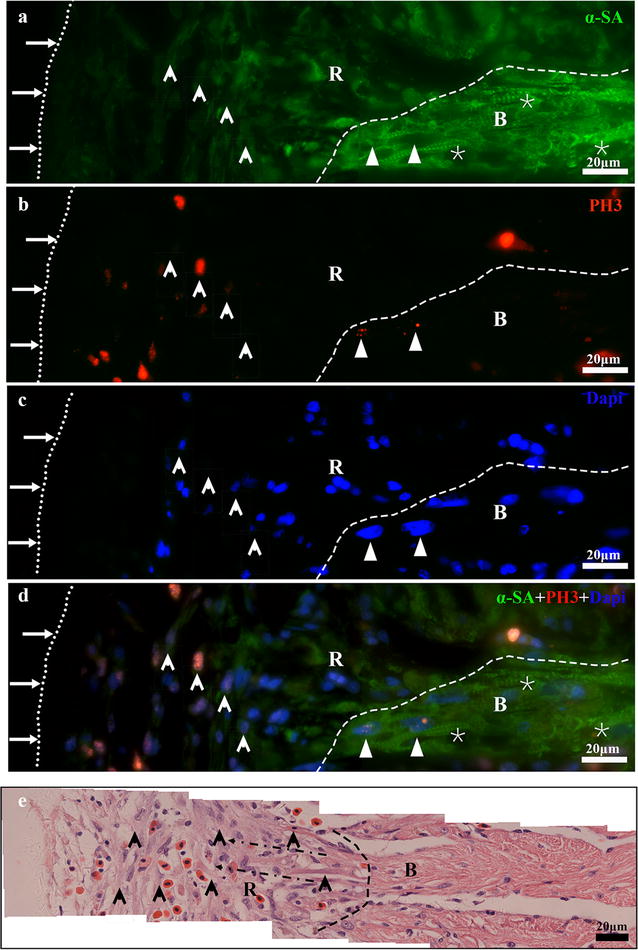
Immunofluorescent staining combined with H&E staining indicating the proliferation of cardiac myocytes during regeneration. a, b Immunofluorescent staining for α-SA+(a) and PH3+ (b) in a same section of the regenerated zone at 8 daar. Nuclei were counterstained with DAPI (c). The overlay image is shown in (d). e H&E staining of section from near dimension of A. Long arrow and small dotted line: Outer surface of the epicardium. Short arrow: Newly regenerated α-SA+/PH3+ cardiomyocytes (a–d) and newly regenerated cardiomyocytes in H&E staining (e). Small triangle: Mature α-SA+/PH3+ cardiac myocytes. Asterisk: mature cardiac myocytes with a regular cross-striated structure. Black arrow with dotted line: Migrating morphology of cardiomyocytes (e). Large white black dotted line: Border of the regenerated zone. R regenerated area, B border area. The results are from three animals
