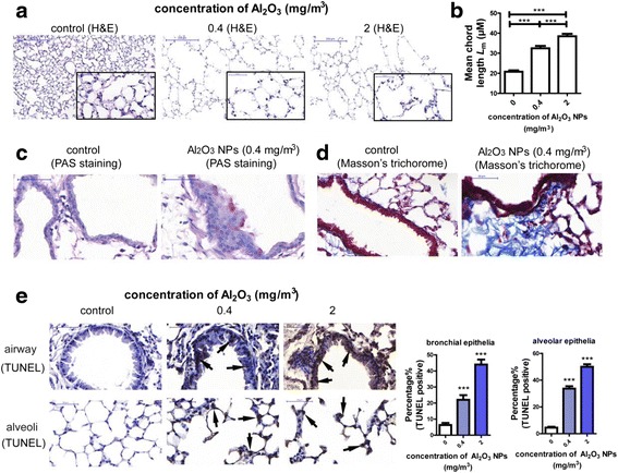
Fig. 2.

Exposure to Al2O3 NPs led to experimental COPD in a murine model. a Representative images of normal alveolar area and emphysema. The images on the right bottom of each panel are magnified (400) of area from the original one. b Lm was significantly increased in Al2O3 NPs-exposed mouse lungs (n = 36, *** P < 0.001). c Representative images of PAS staining, the PAS+ cells suggested hypersecretion of airway epithelial cells (shown by arrows). d Representative images of Masson’s Trichorome staining. Deposit of collagen around airway was stained purple. e Representative images of TUNEL staining and percentage of TUNEL+ cells. The TUNEL+ cells were shown by arrows. (n = 30, *** P < 0.001, compared with control group)
