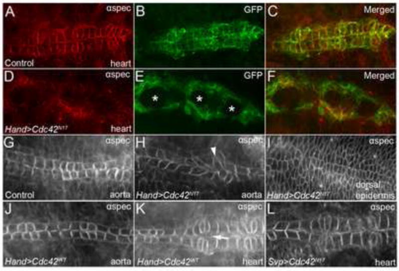
Figure 3. Dominant negative expression of Cdc42 causes severe DV defects.

(A-C) Stage 17 Hand-GAL4,UAS-GFP-moe (control) embryos in whole mount stained with anti-αSpectrin (A) and anti-GFP (B) in the heart region of the DV to show normal CB morphology and alignment at the dorsal midline. (C) is a merged image of (A) and (B). Overexpression of dominant negative Cdc42 (Cdc42 N17) (D-F) in the DV produces large holes (asterisks) within the heart. Anti-αSpectrin staining of the aorta (G-H) reveals abnormal CB morphology (arrowhead) in Cdc42N17 mutants (H) compared to wild type (w1118) aortas (G), albeit no CB contact defects as seen in the heart region (E). (I) Cdc42N17 mutants maintain proper dorsal ectodermal closure, evident by anti-αSpectrin staining. (I) is the same embryo as shown in (D-F) at a different focal plane. (J-K) Overexpression of wild type Cdc42 (Cdc42WT) does not cause any defects in the aorta (J) and results in only minor gaps (arrow) between Svp+ cells in the heart (K). (L) Overexpression of Cdc42N17specifically in Svp+ CBs with Svp-GAL4 results in normal DV closure and CB morphology as apparent by anti-αSpectrin staining. See Table 1 for quantitative data.
