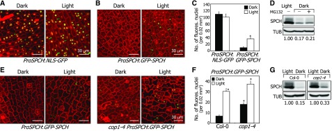
Figure 6.
SPCH Degradation Is Not Associated with Ubiquitin/Proteasome Pathways.
(A) Fluorescence-assisted detection of SPCH promoter activity. Three-day-old ProSPCH:NLS-GFP transgenic seedlings grown in darkness were exposed to white light for 6 h. Confocal images of abaxial epidermal cells of cotyledons were obtained. All scale bars represent the same value. Bars = 30 μm.
(B) and (C) SPCH accumulation in the nuclei of abaxial epidermal cells of cotyledons. Three-day-old ProSPCH:GFP-SPCH transgenic seedlings grown in darkness were exposed to white light for 6 h before obtaining confocal images (B). Bars = 30 μm. Fluorescent nuclei in abaxial epidermal cells were counted (C). Three independent measurements, each consisting of 10 counts, were averaged and statistically analyzed (t test, *P < 0.01). Bars indicate se.
(D) Effects of MG132 on SPCH degradation. Five-day-old transgenic seedlings overexpressing a MYC-SPCH fusion were incubated in darkness for 24 h in the presence of 50 μM MG132 before extracting total proteins from whole seedlings. Immunological detection of SPCH proteins was performed using an anti-MYC antibody. Blots on the membranes were quantitated using ImageJ software.
(E) and (F) Dark-induced degradation of SPCH in cop1-4 stomatal lineage cells. Three-day-old ProSPCH:GFP-SPCH and cop1-4 ProSPCH:GFP-SPCH seedlings grown in darkness were exposed to white light for 6 h before obtaining confocal images of abaxial epidermal cells of cotyledons (E). Bar = 30 μm. All scale bars represent the same value. Fluorescent nuclei in abaxial epidermal cells were counted (F). Three independent measurements, each consisting of 15 counts, were averaged and statistically analyzed (t test, *P < 0.01). Bars indicate se.
(G) SPCH accumulation in the cop1-4 background. Five-day-old 35S:MYC-SPCH seedlings were incubated in darkness for 24 h. Immunological detection of SPCH proteins was performed as described in (D). Blots on the membranes were quantitated as described above.

