Figure 4.
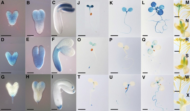
Tissue- and Cell-Type-Specific Expression of DHFR-TS Genes in Arabidopsis.
(A) to (X) Histochemical GUS staining of ProDHFR-TS:DHFR-TS-GFP-GUS Arabidopsis lines.
(A) to (I) Expression of DHFR-TS genes during embryonic development. (A) to (C) DHFR-TS1 expression, (D) to (F) DHFR-TS2 expression, and (G) to (I) DHFR-TS3 expression. Bars = 50 µm.
(J) to (X) Expression of DHFR-TS genes during postembryonic development.
(J) to (L) DHFR-TS1 expression. (J) Three-day-old seedling, (K) 5-d-old seedling, (L) 10-d-old seedling, (M) inflorescence, and (N) flower. Bars = 2 mm in (J) to (L), 1 cm in (M), and 1 mm in (N).
(O) to (S) DHFR-TS2 expression. (O) Three-day-old seedling, (P) 5-d-old seedling, (Q) 10-d-old seedling, (R) inflorescence, and (S) flower. Bars = 2 mm in (O) to (Q), 1 cm in (R), and 1 mm in (S).
(T) to (X) DHFR-TS3 expression. (T) Three-day-old seedling, (U) 5-d-old seedling, (V) 10-d-old seedling, (W) inflorescence, and (X) flower. Bars = 2 mm in (T) to (V), 1 cm in (W), and 1 mm in (X).

