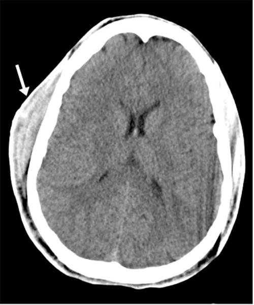
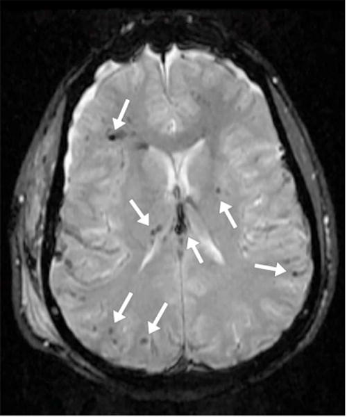
Figure 3.


17 year-old male status post motor vehicle collision. (A) Axial CT image in soft tissue windows demonstrates a large right subgaleal hematoma (arrow) without any evident intracranial abnormality. (B) Axial gradient-recall echo (GRE) MR image in a similar plane demonstrates foci of susceptibility artifact (arrows), presumed to represent microhemorrhage, scattered along the gray white junction, in the right thalamus, in the left putamen, and in the fornix body. SWI has replaced GRE at our institution in most routine imaging protocols. (Images courtesy Liangge Hsu M.D., Brigham & Women’s Hospital)
