Figure 4.


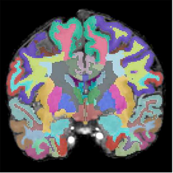
(A) Axial, (B) sagittal, and (C) coronal images of the brain with automatic segmentation label maps generated by FreeSurfer superimposed on T1-weighted high resolution MR images.


(A) Axial, (B) sagittal, and (C) coronal images of the brain with automatic segmentation label maps generated by FreeSurfer superimposed on T1-weighted high resolution MR images.