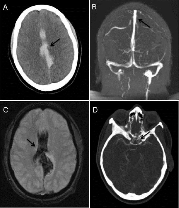
Figure 1.
A) Non-contrast head CT revealing extensive interhemispheric and subarachnoid haemorrhage (arrow). (B) MRV of the Head demonstrating patency of the sagittal sinus in the region of the haemorrhage (arrow) and absence of dural sinus thromboses. (C) MRI brain, hemoflash sequence, corroborating the area of haemorrhage (arrow). (D) CT angiogram of head demonstrating a small, incidental left supraclinoid internal carotid artery aneurysm (arrow). No obvious vasculitic changes or abnormalities in the areas of intracranial haemorrhage seen.

