Figure 2.
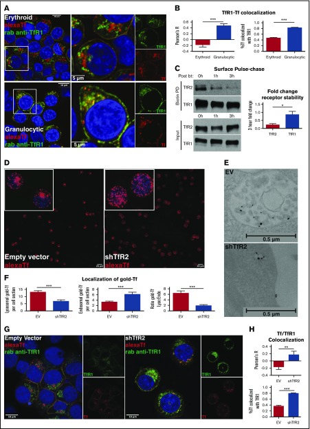
TfR2 is required for the lysosomal trafficking of transferrin. (A) Fluorescence microscopy for colocalization of Alexa Fluor 594-transferrin with TfR1 in erythroid progenitors (top left: confocal image with 63× oil objective; top right: inset of image depicting higher magnification) and granulocytic progenitors (bottom left: confocal image with 63× oil objective; bottom right: inset of image depicting higher magnification). Yellow denotes merge between separate red and green channels. (B) Colocalization of Alexa Fluor 594-transferrin and TfR1, as measured by Pearson’s correlation coefficient (left) and percentage of Alexa Fluor 594-transferrin+ vesicles co-occupied by TfR1 (right) (number of cells analyzed = 27-58 per group; ***P < .001). (C) Immunoblot of surface-biotinylated proteins from progenitors in erythroid medium with diferric transferrin, with indicated duration of culture in hours postbiotinylation (left). Densitometry for TfR2 vs TfR1 fold decline over the course of 3 hours postbiotinylation (right) (n = 4; *P < .05). (D) Fluorescence microscopy of human erythroid progenitors transduced with lentiviral shRNA constructs and subjected to Alexa Fluor 594-transferrin uptake (confocal image with 10× objective, insets with 63× objective). (E) Transmission electron microscopy of erythroid progenitors transduced with lentiviral shRNA constructs and subjected to 10 nm gold-conjugated transferrin uptake highlighting MVB/lysosomal accumulation (top) and endosomal accumulation (bottom) of transferrin particles. (F) Summary of transmission electron microscopy studies as in panel E showing the number of lysosomal transferrin particles per cell section with indicated lentiviral shRNA transduction (left); number of endosomal transferrin particles per cell section with indicated lentiviral shRNA transduction (middle); and ratio of lysosomal to endosomal localization of transferrin (right) (number of cells counted = 44-45 per group; ***P < .001). (G) Fluorescence microscopy for colocalization of Alexa Fluor 594-transferrin with TfR1 in erythroid progenitors transduced with lentiviral shRNA constructs (confocal image with 63× oil objective). Yellow denotes merge between separate red and green channels. (H) Colocalization of Alexa Fluor 594-transferrin and TfR1 as measured by Pearson’s correlation coefficient (top) and percentage of Alexa Fluor 594-transferrin+ vesicles co-occupied by TfR1 (bottom) (number of cells analyzed = 12-23 per group; **P < .01, ***P < .001).

