Figure 5.
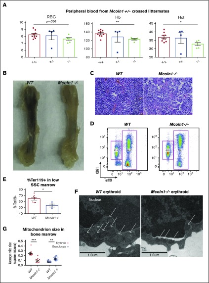
The lysosomal iron channel Mucolipin-1 contributes to erythropoiesis and mitochondrial integrity. (A) Circulating red blood cell counts, hemoglobin concentration (Hb), and hematocrit (Hct) in littermate adult wild-type, Mcoln1+/−, and Mcoln1−/− mice (n = 9 per group; *P < .05, **P < .01). (B) Femur images from adult wild-type and Mcoln1−/− mice. (C) Light micrographs of hematoxylin and eosin–stained bone marrow from adult wild-type and Mcoln1−/− mice (40× objective). (D) Flow cytometry of wild-type and Mcoln1−/− bone marrow erythroid progenitors to assess erythroid differentiation. (E) Summary of flow cytometry studies as in panel D, showing decrease in percentage of Ter119+ cells in low SSC-gated Mcoln1−/− bone marrow (n = 4-8 per group; *P < .05). (F) Transmission electron microscopy of bone marrow depicting marrow erythroid progenitors from adult wild-type (left) and Mcoln1−/− (right) mice; arrows denote mitochondria. (G) Ultrastructural quantitation of mean mitochondrial size in erythroid and granulocytic progenitors from wild-type and Mcoln1−/− bone marrow as in (F) (number of cells analyzed = 10 per group; ***P < .001; **P < .01).

