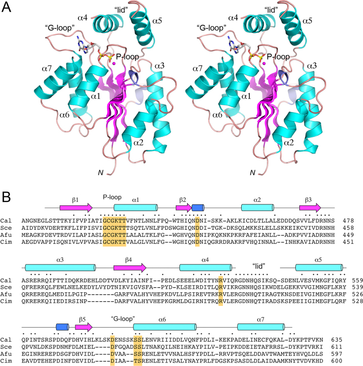
Figure 2.
Structure of the CalTrl1 kinase domain. (A) Stereo view of the kinase tertiary structure, depicted as a ribbon model with magenta β strands, cyan α helices (numbered sequentially), and blue 310 helices. The N-terminus (Leu407) is indicated by N. The GDP in the phosphate donor site is rendered as a stick model. Mg2+ is depicted as a magenta sphere. The phosphate-binding P-loop and guanine-binding ‘G-loop’ are indicated. (B) Secondary structure elements (colored as in panel A) are displayed above the CalTrl1 KIN primary structure, which is aligned to the primary structures of the KIN domains of S. cerevisiae (Sce), A. fumigatus (Afu) and C. immitis (Cim) Trl1. Positions of amino acid side chain identity or similarity in all four proteins are indicated by dots above the Cal sequence. Gaps in the alignment are indicated by dashes. Conserved P-loop, aspartate general acid, lid, and G-loop elements are highlighted in gold shading.

