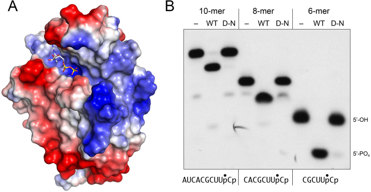
Figure 5.
Surface electrostatics and minimized 5′-OH RNA substrates for the KIN domain. (A) A surface electrostatic model of the KIN domain was generated in Pymol. GDP in the phosphate donor site is depicted as a stick model. (B) Reaction mixtures (10 μl) containing 50 mM Tris–HCl (pH 7.5), 50 mM NaCl, 2 mM DTT, 10 mM MgCl2, 100 μM GTP, 20 nM 32P-labeled HORNA3’p of varying length as specified (depicted at bottom, with the 32P-label indicated by •) and either 100 nM wild-type KIN domain (WT), D445N mutant (D–N), or no enzyme (–) were incubated at 22°C for 20 min. The labeled RNAs were resolved by urea–PAGE. An autoradiogram of the gel is shown. The positions of the 6-mer 5′-OH RNA substrate and 5′-PO4 RNA product are indicated on the right.

