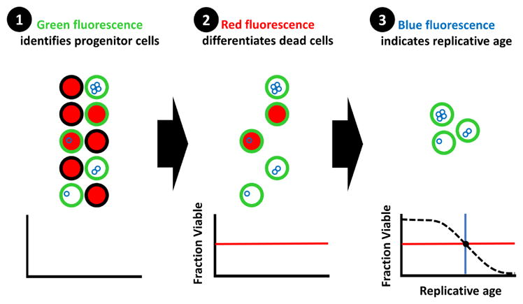
Fig. 1. Workflow of High-Life experiments.

A visual depiction of the work-flow for High-Life experiments. The progenitor cell population of interest is persistently labeled with a green NHS-Ester fluorescein conjugate, which asymmetrically segregates to the mother cell during division. The fraction of cells viable within the progenitor population is then determined using propidium iodide; live cells exclude the red dye. Finally, the replicative age of viable progenitor cells is measured using wheat germ agglutinin conjugated to a blue fluorophore, which labels bud scars left behind with each division. A complete lifespan curve can be constructed using serial measurements taken over the course of 2–3 days.
