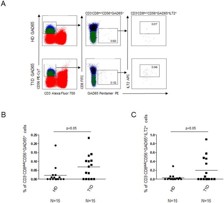
Fig 3. Increased percentages of CD3-CD8dullCD56+ and CD3-CD8dullCD56+ILT2+GAD65 AA 114–122 pentamer reactive cells in T1D patients.
Relative percentages of CD3-CD8dullCD56+ and CD3-CD8dullCD56+ILT2+ GAD65 AA 114–122 pentamer reactive cells in T1D patients vs healthy controls after GAD65 peptide stimulation. (A) Representative experiment showing the flow-cytometry gate strategy; (B) Summary of results of CD3-CD8dullCD56+ GAD65 AA 114–122 pentamer reactive cells obtained in 15 healthy donors (circle dots) and 15 T1D patients (square dots); (C) Summary of results of CD3-CD8dullCD56+ ILT2+ GAD65 AA 114–122 pentamer reactive cells. Horizontal bars, average values are reported.

