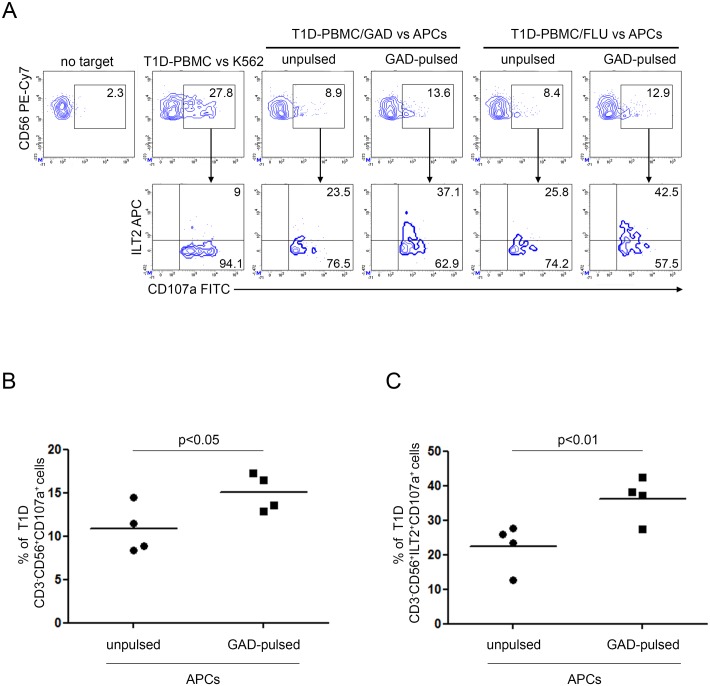
Fig 5. Increased susceptibility of GAD65 AA 114–122 peptide-pulsed APCs to T1D NK cell-mediated recognition associates with NK cell-ILT2 expression.
Degranulation of CD3-CD56+ILT2+ NK cells of PBMC from T1D patients, expanded with GAD65 AA 114–122 or FLU peptides, measured as CD107a cell-surface expression following stimulation with APCs, either left unpulsed or GAD65 AA 114–122 peptide-pulsed. (A) A representative experiment out of two performed is shown. K562 cells were used as positive control. The percentage of CD3-CD56+CD107a+ NK cells (upper panel) and CD3-CD56+CD107a+ILT2+ (lower panel) is indicated for each condition. (B) Summary of CD3-CD56+CD107a+ and (C) CD3-CD56+ILT2+CD107a+ NK cell percentage of four T1D PBMC, expanded with GAD65 AA 114–122 or FLU peptides, following stimulation with APCs, either left unpulsed (circle dots) or GAD65 AA 114–122 (GAD65)-pulsed (square dots); horizontal bars, average values are reported.

