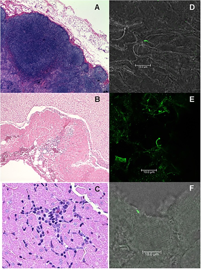
Fig 5. Histopathology and B. burgdorferi (Bb) within tissues of persistently-infected macaques.
At 12–13 months post-infection, necropsy tissues were analyzed for evidence of tissue pathology. Frozen sections of affected tissues were then stained with fluorescently-labeled Borrelia-specific polyclonal antibodies. Shown here is: axillary lymph node hyperplasia and histiocytosis-200X (A) and Bb antigen (D) in untreated animal IN16; cervical spinal cord, focal inflammatory lesion-200X (B) and Bb antigen (E) from treated animal IH11; and a focal area of mononuclear inflammation in the myocardial interstitium-1000X (C) and Bb antigen (F) from untreated animal IN05.

