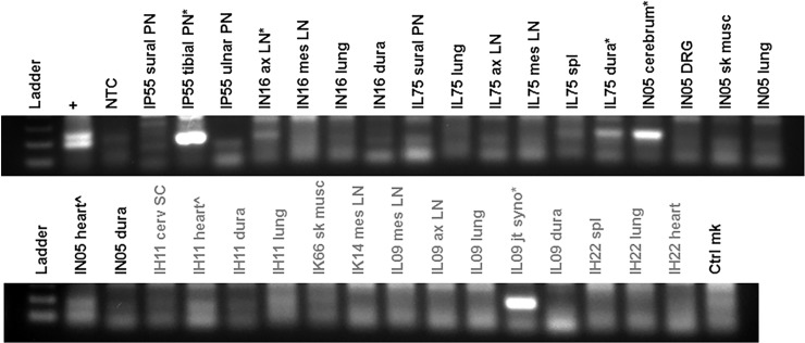
Fig 6. Amplification of B. burgdorferi-specific DNA in tissues showing histopathology.
Tissue DNA was subjected to amplification of a region within the 5s-23s ribosomal genes using a nested set of primers. The positive control (+) consisted of B31.5A19 DNA and negative controls were no template control (NTC) and uninfected monkey skin tissue DNA (Ctrl mk). Tissues tested included peripheral nerves (PN), axillary (ax) and mesenteric (mes) lymph nodes (LN), lung, meninges/dura mater (dura), spleen, cerebrum, skeletal muscle (sk musc), heart, spinal cord (SC), and joint synovium (jt syno). Clear positives are marked with (*) and potential positive results are marked with (^). The treated monkeys are labeled with grey text.

