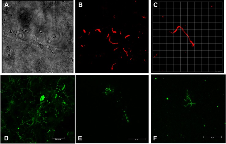
Fig 7. Spirochetes identified from in vivo culture of monkey heart tissue.
The DMC-cultured heart tissue was placed in BSK and back to in vitro culture for 1 week. The culture pellets were then stained with a combination of anti-OspA and anti-OspC monoclonal antibodies, followed by a red detection antibody (Panels A,B,C) or with anti-OspA followed by a green detection antibody (Panels D,E,F). Shown are differential interference contrast (Panel A) and fluorescent detection (Panels B and C) of cultures stained with monoclonal antibodies. Panels A and B: animal IN05 (untreated); Panel C: animal IH22 (treated). Panel D is a positive control B. burgdorferi culture; Panels E and F are stained specimens from heart cultures of monkeys IK14 and IL09 (both treated), respectively.

